3D Printing Unwraps Mummy Mystery


Latest News
September 12, 2016
3D scanning and printing are being used in medical applications to help doctors plan for complex surgeries, to help create custom-fit prosthetics, and to manufacture medical implants. But that same technology can be used to help archaeologists study “patients” that are beyond treatment – like a 2,000-year-old mummy.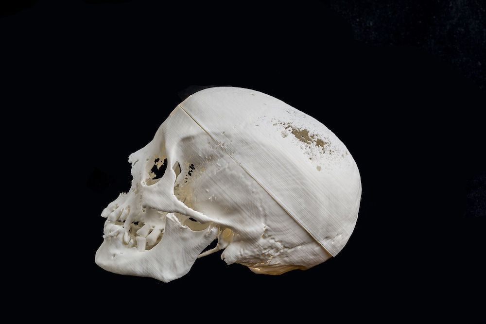
The University of Melbourne in Australia leveraged 3D printing to study an Egyptian mummy’s head, reconstruct her face and even point to a disease that may have factored in her death. They were able to do this all without compromising the original remains.
A museum curator discovered the previously undocumented mummy head in the University’s collection during an audit, and ordered a computed tomography (CT) scan of the remains to determine their condition.
Staff in the University’s Department of Anatomy and Neuroscience worked with imaging specialist to turn the scans into a 3D-printed skull. It took 140 hours to print the skull on a consumer-style 3D printer.
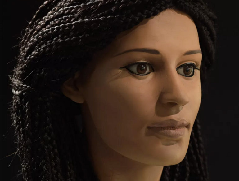
 A forensic sculptor used the 3D printed skull to recreate the mummy’s face. Image: University of Melbourne
After studying the characteristics of the skull, they determined the mummy belonged to a woman (whom they dubbed Meritamun), and that she was roughly 25 years old. The bandaging and embalming techniques indicate the mummy is Egyptian, and may be 2,000 years old. Radiocarbon dating will be used to more precisely determine the age of the mummy. Sculptor Jennifer Mann recreated the mummy’s face.
The skull also indicated that Meritamun suffered from dental abnormalities and severe anemia. The anemia would have caused swelling of bone marrow, which would explain the thinning scientists observed on the top of her skull.
“It is quite fascinating that we did all of this without destroying the specimen in any way, and that is important from a museum curatorial point of view,” said Varsha Pilbrow, a biological anthropologist in the University of Melbourne’s Department of Anatomy and Neuroscience.
The ability to scan and print copies of items in the University’s specimen collection has made it easier for students and researchers to work with the collection. “We can now replicate specimens with really interesting pathologies for students to handle and for virtual reality environments without ever touching the specimen itself,” says Gavan Mitchell, the imaging technician at the Department of Anatomy and Neuroscience.
It’s still unclear how the mummy came to be in the museum’s collection. In a Live Science article, staff speculated that former head of the anatomy department, Frederic Wood Jones, may have brought the mummy with him from Egypt after conducting archeological work there in the 1920s The mummy is housed at the Harry Brookes Allen Museum of Anatomy and Pathology.
Source: Live Science and the University of Melbourne
Subscribe to our FREE magazine, FREE email newsletters or both!
Latest News
About the Author
Brian Albright is the editorial director of Digital Engineering. Contact him at [email protected].
Follow DE